Fabrice MoretIng. Dipl. ETS/HES
|
Affiliation:
Univ. of Freiburg, Ophthalmology
|
|---|
Home
CV
Research *
Teaching
Publications
Past projects
Current research
In glaucoma, the blood flow feeding the retina is impeded. It remains to be determined if these alterations are causative of the disease or if they are side effects. To support this investigation, a better understanding of the healthy physiology as well as better tools are required. Our research is geared toward these three points.Specifically, we work on new optical techniques for retinal imaging in humans. Our approach combines both image recording techniques and image processing. The recording techniques aim at collecting quantitative images, a prerequisite to most image processing to be applied later on. The image processing then aims at removing the remaining unwanted variations to offer clean sequences. Finally quantitative parameters, e.g. vessel diameters, can be measured over time and help our understanding of the eye's physiology.
Our techniques are non-invasive, requiring no contact and no injection of contrast agent and thus very comfortable for patients. They also present an interesting potential: installed clinical equipment can be upgraded with little or no additional hardware, allowing fast and affordable transfer from research to patient care.
Various other ocular or systemic diseases relate to blood flow alterations in the eye: diabetes mellitus, hypertension, and intracranial hypertension to name a few. As our techniques and new understanding of the relevant physiology overlap with questions relating to these diseases, our investigations naturally encompass them as well.
1. Pulsation of the retinal vessels
The pulsations of retinal vessels are of interest for understanding ocular blood flow and diagnosing. For instance when measured before and after a vascular segment, the differential pulsation amplitude allows quantification of its resistance to the flow. The local pulsation of the veins is also of interest: first, it as is influenced by the intracranial pressure, it may offer a way to measure it non-invasively and, second, the nature of its relationship to glaucoma remains to be clarified and may offer clues to its aetiopathology.The observation of fine pulsations is often impossible by direct observation and remains difficult even on recorded movies. Visualization and measurements of these pulsations are strongly limited by noise, eye movements and many other factors. We developed an image processing technique to address these limitations.
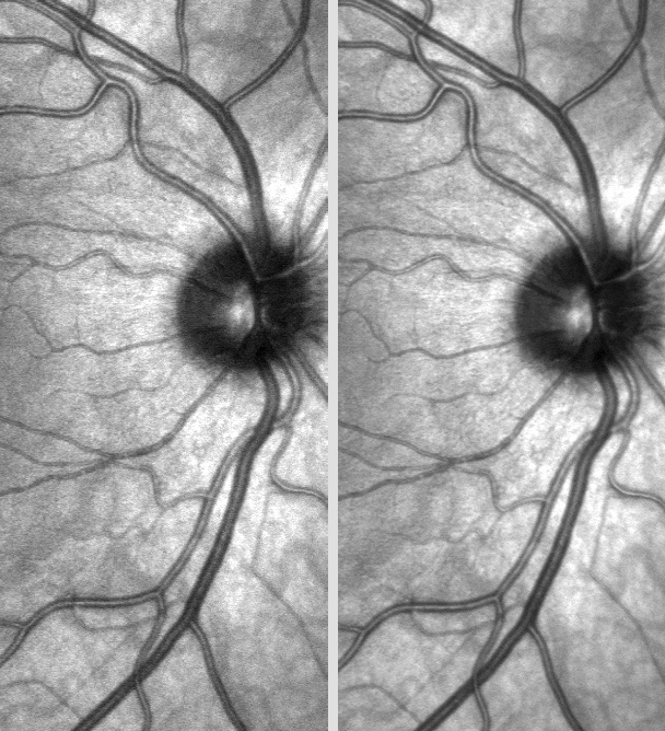
Movies of the retina of a healthy subject showing arteries and veins (respectively the
brighter and darker lines) radiating around the optic nerve head (the dark spot),
the region where the nerve fibres exit the eye globe and proceed to the brain.
o The left movie was acquired with a clinical imager and shows the typical
micro-movements the eye performs when one is asked to gaze steadily at a point.
o The right movie shows the result of the image processing: the pulsations of the
arteries (look at the vessels bending) and veins (e.g. just above the optic nerve head)
are now clearly visible.
See more movies here,
and read
more
(Moret et al. IOVS 2011)
2. Functional imaging
Not only is the blood flow impeded in glaucoma, but also the mechanism adjusting it to the level of neural activity in the retina (neurovascular coupling). This project aims at developing a technique to image the vascular response to a visual stimulation of the retina, and to investigate the origin of the impairment.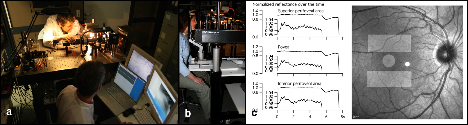 Successful initial development in primates whereupon transferred to humans: Two instruments
have been developed and equipped with a visual stimulation (a, b). Pilot experiments
have confirmed the recordability of the visually evoked vascular signal (c). Still, the
signals have proven to be strongly dependent on the ambient conditions and subjects. We are
now working to make our experimental system robust enough to allow recording in >10 subjects
and start conducting pilot clinical testing.
Successful initial development in primates whereupon transferred to humans: Two instruments
have been developed and equipped with a visual stimulation (a, b). Pilot experiments
have confirmed the recordability of the visually evoked vascular signal (c). Still, the
signals have proven to be strongly dependent on the ambient conditions and subjects. We are
now working to make our experimental system robust enough to allow recording in >10 subjects
and start conducting pilot clinical testing.3. Quantitative access to the retina
Acquiring retinal images for clinical purposes requires specialized skills yet can be performed routinely: a contrast sufficient for visual examination is the key quality metric. One the contrary, acquiring images for visualizing changes over time using post-hoc image processing requires a specific type of fundus imaging: quantitative imaging. Here the key parameter is not the contrast anymore but the stability of the images, stability meaning that the measured pixels' intensities only fluctuate over a range of a few percent. As the eye is incredibly dynamic, a wealth of parameters must to be taken into account during image acquisition.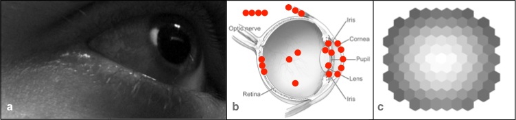 To achieve quantitative imaging, we first identified >20 factors involved (b) and
iteratively addressed them, either by instrumental developments or by
adjusting the recording methodology. Our system now routinely yields high quality
image sequences of >30 seconds and is still continuously being improved.
The need for quantified access to the retina is also true for the visual stimulation.
Here we developed a simple method to calibrate the spatial intensity of a visual
stimulus when displayed in the eye by an instrument instead of simply being presented over
a screen (c).
To achieve quantitative imaging, we first identified >20 factors involved (b) and
iteratively addressed them, either by instrumental developments or by
adjusting the recording methodology. Our system now routinely yields high quality
image sequences of >30 seconds and is still continuously being improved.
The need for quantified access to the retina is also true for the visual stimulation.
Here we developed a simple method to calibrate the spatial intensity of a visual
stimulus when displayed in the eye by an instrument instead of simply being presented over
a screen (c).