Fabrice MoretIng. Dipl. ETS/HES
|
Affiliation:
Univ. of Freiburg, Ophthalmology
|
|---|
Home
CV
Research
Teaching
Publications
Past projects *
Crisis syringe pump – COVID-19
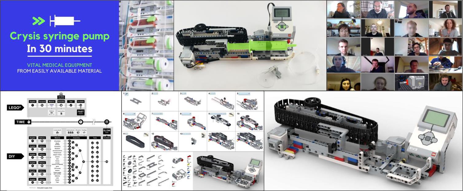
To palliate the lack of syringe pumps for ventilated COVID-19 patients, during the
early phase of the pandemic outbreak, an international group of >30 engineers,
scientists, nurses, doctors, industry experts and volunteers gathered to develop
a crisis pump, to be built in 30 minutes, out of a single Lego set.
Following a statement in the press by the head of Intensive Care Unit of the Geneva
University Hospitals, together first with a compact team, I initiated the project
and quickly backboned it on the Swiss authorities hackathon
VersusVirus.ch.
The project was then powered by the EU hackathon
EuVsVirus.org
for which we were selected as finalists. The incredible team assembled did managed the feat
of designing a proof of concept hardware apparatus exclusively through video
conferencing.
We are now preparing
publications for documenting this agile reaction to a crisis, and further
disseminating the device, e.g. as an open pump for medical and biosciences
research laboratories.
See
more
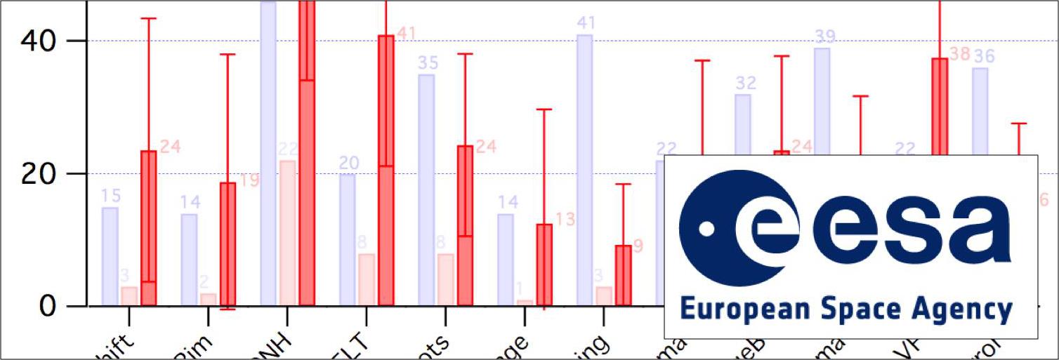
Figure for the Solidarity study – COVID-19
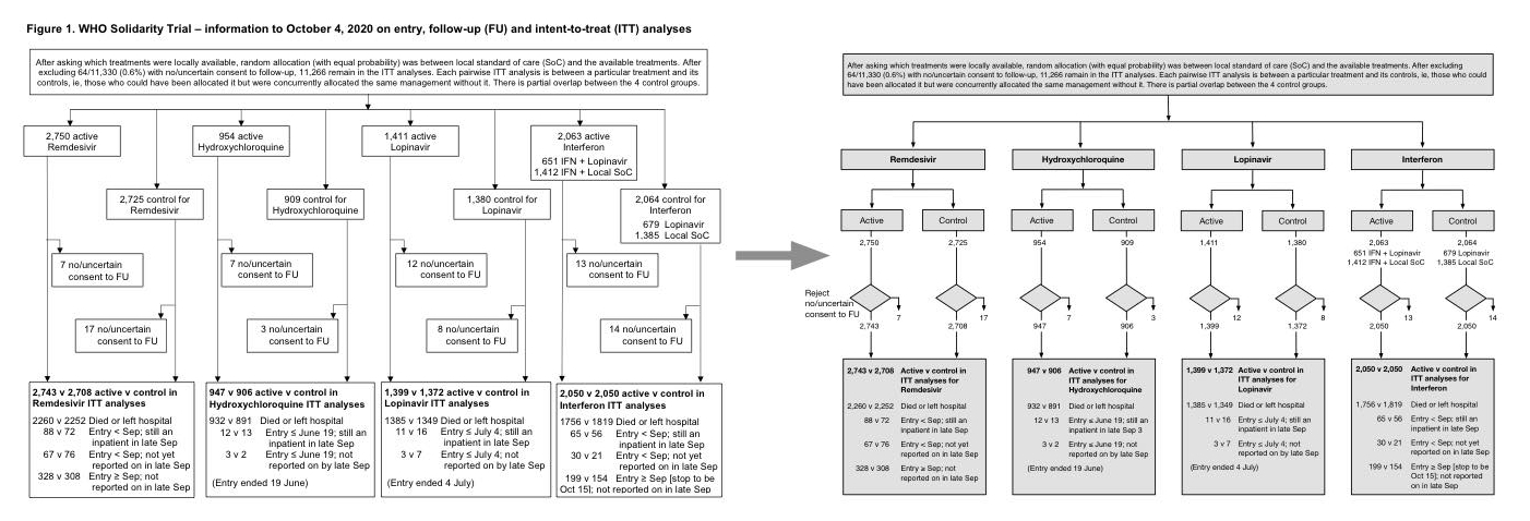
The Solidarity study tested repurposed antiviral drugs for Covid-19. I volunteered a redo
of the preprint figure illustrating the methodology of the study (four branches with
controls).
Visualizations generally benefit from understanding of the Gestalt principles, and
from seeking the optimal way to code information such that a single glance
provides clear perception and insights of complex concepts (“inference at a glance”).
Applications of visual engineering range from representing data, methodologies,
engineering schemas of complex systems, to the creation of visual interfaces for
demanding environments (e.g. medicine or aeronautics).
I am open to contributing to scientific projects on public health with community
benefits. (Agile process: You email me, I tell you immediately if it is possible. I do
not ask for authorship or attribution.)
Data visualization tool for distributions
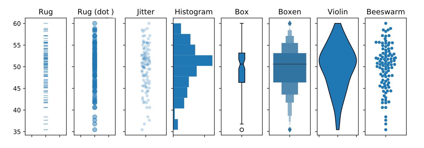
Data visualization is a critical step for exploratory analysis and for
communication. The representation of data distributions, one of the key
visualization, benefited from the introduction of markedly improved tools
over the last years. Yet no single tool combines the desired features of being
totally intuitive, free of arbitrary parametrization, representing the raw data,
and being free of visual biases.
I am currently developing a tool addressing these needs. I reviewed the state of
the art and implemented a test bench to assess the various visualization tools
with different data configurations. The final tool is developed as a Python
library, to be used as a complement to existing visualization libraries.
Skeletonizing C elegans
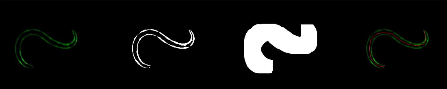
C elegans have short life cycle and are simple to handle, thus allow high
throughputs modeling for life sciences. Here I developed a simple image processing
pipeline to retrieve a clean backbone from images of worms, to later allow extra
processing for observation and quantification.
From the raw fluorescence image, the ROI including the specimen is identified and
framed (a), the image is thresholded, inflated to fill
segments w/o fluorescence, skeletonized, skeleton pixels are sorted to form a
continuous segment using graph theory, then a parametric function, and finally a
B-spline is interpolated from
the skeleton. The processing is done with Python, openCV, SciPy and Scikit-learn
(Data: Nagi Bioscience).
Astronaut's health data analysis – ESA
Long-term stays in microgravity, such as in the International Space Station,
lead to ocular alterations for the astronauts. This is identified
as a major hurdle to overcome to open the way for the manned exploration
of Mars.
Extensive studies have been conducted to characterize ocular alternations,
to understand their etiology, and to device countermeasures. Here I proposed a
new statistically analysis to provide insights on subgroups stratification of
a large cohort of astronauts and I generated state-of-the-art data visualizations,
for the ESA medical board.
Color Doppler Imaging – Vascular signals extraction
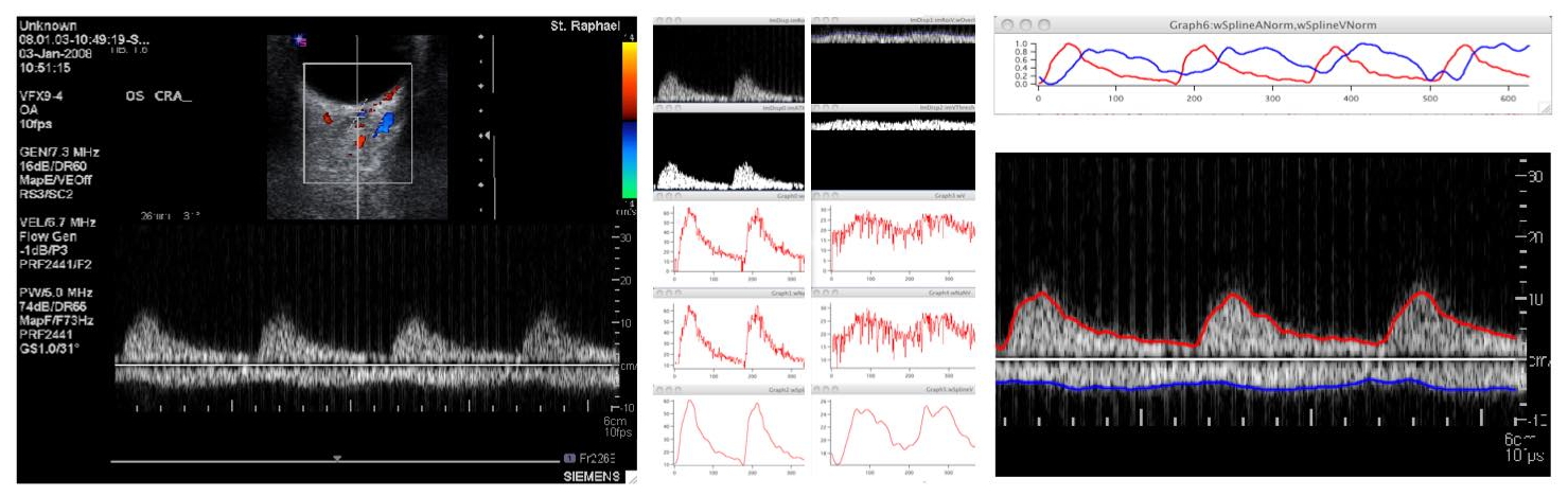
Color Doppler Imaging is one of the various modalities to image and measure the
vascular activity in the eye. In a clinical setting at the University of Leuven,
the device did not provide the envelopes of the arterial and venous flow signals.
An image and signal processing pipeline was established to extract these signals
(as splines) from the screen images, to allow further visualization and
quantitative analysis. In collaboration with Prof. Abegão Pinto from the Centro
Hospitalar Lisboa Norte, University of Lisbon.
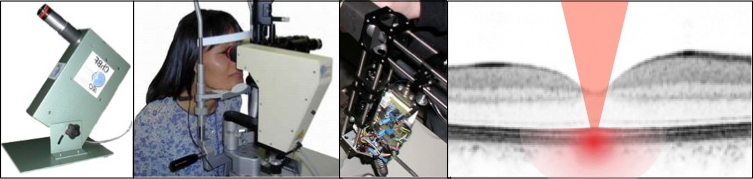
Characterization of human corneal grafts
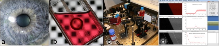
Cornea transplantations (a) are performed to restore or improve vision in patients
with damaged or diseased corneas. Ninety-five percent of the patients recover sight
after
this surgery. To investigate the origin of the limited visual acuity in the
remaining 5%
and in an attempt to predict the surgical outcome, we developed an optical bench to
characterize the optical quality of the ex-vivo grafts.
The instrument measured the Modulation Transfer Function of grafts still immersed in
their nutrient baths (b), using a real-time slanted edge super-resolution algorithm
(c,d). A swelling of the ex-vivo cornea due to deficiencies in fluid out-pumping occurs
rapidly and generates folds impeding the optical quality. Meanwhile, once
transplanted,
these folds resorb. Measurements in five grafts showed that the initial
swelling is critically important compared to post-operative resorption, and thus
representative metrics of the cornea's optical quality cannot be acquired under
the tested
conditions. This project was performed at the Dept. of Ophthalmology, Univ. of
Freiburg, with Dr. Sonja Heinzelmann and Dr. Philip Maier, in 2009–10.
Retinal blood flow in microgravity
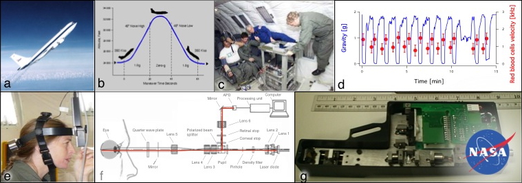
One third of astronauts report changes in their vision in space. To investigate the
etiology of these changes, we measured choroidal blood flow in microgravity.
The choroid is
one of the two vascular systems supplying the retina and has been hypothesized to
engorge as the balance of body fluids shifts toward the head.
Measurements conducted on-board NASA's KC-135 airplane (a-c) have shown that
choroid hemodynamics react to transient high- and zero-gravity (d) and thus that
regulation mechanisms are insufficient to compensate for the induced ocular
perfusion
pressure changes. This confirms the choroid as potentially involved in the reported
losses of visual acuity and warrants further investigations.
Hemodynamic measurements were performed using a Laser Doppler flowmeter redesigned by
FM for flight constraints (e-g), based on the prototype developed at IRO
under
the supervision of Prof. Geiser and Prof. Riva who pioneered the technique. The optical
core of the instrument was robotically assembled with Dr. Lambelet using his Optical
SMD technology (BrightPower SA, an EPFL spinoff). Dr. Petrig
from Oculix SA provided the real-time Doppler Spectrum FFT processing unit.
Prof. Ansari from NASA/GRC supervised and hosted the project in 2001–02.
FM was supported by the Swiss Academy of Engineering Sciences.
Multifocal electroretinograms analysis
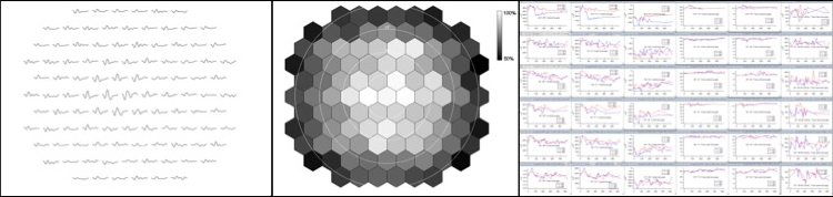
mfERG (see project below) data are highly multidimensional: A simple exam implies up to
103 hexagons each generating >100-points signals (a, b). A study involves exams repeated
over time, on multiple subjects and finally the
analyses require pooling responses from rings, hemi-fields and various other groupings.
There is no commercial software geared toward such analyses hence a tool has been
designed.
Software written by Prof. Bach served as the starting point. The new version accommodated
more hexagons and featured various graphical user interfaces separated from core
functions.
This allowed both fast single record visualization (a, b) and batch processing of massive
study datasets by command lines (c). The signal processing is organized as a
linear pipelined structure, to favor maintenance and signal processing fast prototyping.
This project was conducted in 2006–08 with Profs. Bach and Kremers.
Fundus-controlled multifocal electroretinogram
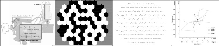
The multifocal electroretinogram (mfERG) is an exam of the retina where the
electrophysiological response to a visual stimulus is recorded by means of electrodes
lying on the sclera. The mfERG's particularity is to provide an array of responses,
and thus spatial information, as a complex patterned stimuli is used.
In the elderly with altered central vision, children or anesthetized animals, stimulus
centering cannot rely on the subject's skills or collaboration. This project aimed at
alleviating this need by providing a fundus-control, i.e. a visualization of the retina,
allowing the operator to perform the alignment himself.
A fundus camera was converted for digital near-infrared imaging and a visual stimulator
was fitted onto it. The system was assessed in primate experiments and results showed
good matching with conventional mfERG systems. This project was conducted at the
Novartis Institutes for Biomedical Research in 2003–05 with Dr. Doelemeyer and
Prof. Kremers.
Eye-tracked flowmeter
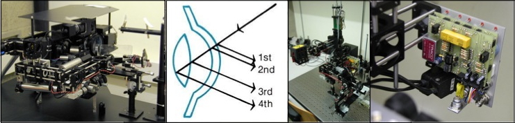
The blood flow in the back of the eye is thought to be involved in many ocular diseases
and thus requires proper investigatory tools. As the back of the eye
is a highly heterogeneous structure, blood flow measurements have to target fine elements,
e.g. vessels approximately 100 µm wide. Pinpointing such vessels is difficult as the eye
is
constantly moving and as these movements may have larger amplitudes than the vessel's
dimensions. To address this limitation a research-grade instrument correcting for
movements in real-time was designed by Prof. Riva and Dr. Mendel. To allow
rapid measurement in elderly patients with less efficient gaze fixation,
a new design was proposed by Prof. Geiser. This design merged the previously
separated laser illumination and collection path in a common confocal scheme, thus
simplifying the operation of the instrument.
Starting from the initial instrument and the optical simulation of the new scheme,
FM implemented the new confocal eye-tracked optical system and a fundus imaging system
onto
a dual-Purkinje-image eye tracker. This work was performed at IRO in 1999–2001.
Visual stimulator for neurovascular coupling investigations
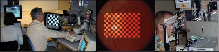
Not only is the blood flow impeded in glaucoma, but also the mechanism adjusting it to
the level of neural activity in the retina (the neurovascular coupling). To measure both
components a Laser Doppler flowmeter measuring the blood flow has to be combined with a
pattern electroretinogram system (pERG)(fig. a). The pERG records the bioelectrical
responses of specific retinal cells utilizing a reversing checkerboard as visual
stimulation.
The aim of this project was to add a stimulation module onto a flowmeter, allowing
projection of an alternating checkerboard onto the retina (b), this was in order to
simultaneously measure the blood flow and the electrophysiological responses.
Using custom optics, a video projector was fitted on a fundus camera equipped
with a flowmeter (c). This project was performed at IRO in 1999–2000, under the
supervision of Prof. Geiser and destined to Prof. Riva and Prof. Falsini.
Choroidal flowmeter
Ocular blood flow is altered in many ocular or systemic diseases. Prof. Riva
established a method whereby the movement of the red blood cells can be measured using
the Doppler effect that occurs when laser light encounters moving cells.
Based on Riva's instrument, Prof. Geiser proposed limiting the measurement ability of
the initial system to the sub-fovea region, where the choroid can be reached optically
(d),
thus alleviating the need for the complex retinal imaging otherwise required and
yielding a more specialized but simple and user-friendly instrument. This instrument
was the first step toward the miniaturized and wearable flowmeter used in the
microgravity project (see above).
The project's aim was to reassemble the prototype instrument (a) into a robust and
slit-lamp mounted version (b, c). This project was performed at IRO in 1999–2000,
under the supervision of Prof. Geiser.
Micro-scanner for barcode reading
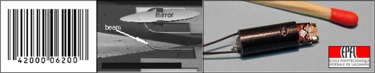
Monolithic silicon integrated optical micro-scanners were developed by Dr.
Schweizer and Prof. Renaud at EPFL (central picture) to offer a low-cost, robust
and small alternative to rotating or oscillating mechanisms then in use in barcode
scanners. These scanners consisted of a 800 x 500 µm mirror supported by thermal-bimorph
actuators and fabricated using a process compatible with IC fabrication
techniques.
Prof. Geiser, Dr. Schweizer and FM designed and assembled an integrated head (right)
with laser delivery from an optical fiber and a grin lens, and measured the planarity of
the mirror using a custom-built test bench. Scanners of this type became industry
standards. The project was conducted in 1999–2000 at the HEVs and EPFL.
Science promotion in classrooms
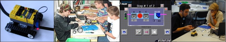
Science and technology are not attractive enough for youngsters, resulting in a
country-wide shortage of engineers in training. As a contribution to reverse this trend,
promotional activities were held in secondary education in the form
of robotic laboratories.
These laboratories where pilot activities with volunteer teachers aimed at
establishing pedagogical materials and teaching formats. Equipment
such as microprocessor units, computers with suitable programming environments,
and Lego technic kits with motors and sensors were provided in classrooms and teachers
trained in robotics. An engineer was present to interact in the classes, presenting their
work and providing hands-on help with robot design. Students were organized in small
groups of three or four and assigned to build various robots such as mobile line
followers.
These activities were intended to promote imaginative and creative use of techniques,
development of observational skills, and use of the experimental approach.
The project was sponsored by the Studying Incentive Commission of the
Swiss Academy of Engineering Sciences. FM contributed to the preparation of
pedagogical materials and animated classes in Bex, Thun and Sion, in 2000–01.